
 https://doi.org/10.4081/cc.2026.15934
Divergent acute release of CGRP and PACAP from mouse spinal cord tissue: implications for migraine neurobiology
All claims expressed in this article are solely those of the authors and do not necessarily represent those of their affiliated organizations, or those of the publisher, the editors and the reviewers. Any product that may be evaluated in this article or claim that may be made by its manufacturer is not guaranteed or endorsed by the publisher.
Accepted: 16 October 2026
Published: 30 October 2026
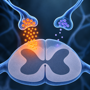
Calcitonin gene-related peptide (CGRP) and pituitary adenylate cyclase-activating polypeptide (PACAP) are both implicated in migraine, but whether they undergo similar acute release from primary sensory terminals remains uncertain. We compared stimulus-evoked CGRP-like immunoreactivity (CGRP-LI) and PACAP-like immunoreactivity (PACAP-LI) in superfused mouse spinal cord tissue. Spinal cords from 14 male C57BL/6 mice were pooled and distributed among six superfusion chambers per stimulus. After two baseline fractions, tissues were exposed to capsaicin (10 μM) or high K+ (40 mM KCl), followed by a post-stimulation fraction. Capsaicin and high K+ produced marked CGRP responses, with mean stimulation-minus-baseline increases of 44.89 and 47.19 pg/mg, respectively, both Holm-adjusted p<0.001. In contrast, PACAP responses were −0.12 and 0.009 pg/mg, with Holm-adjusted p=0.742 and p=0.888. The geometric mean stimulation-to-baseline responses were 23.23-fold and 18.57-fold for CGRP, compared with 0.86-fold and 1.00-fold for PACAP. The corresponding CGRP-to-PACAP ratios of fold responses were 26.99 and 18.51. These findings show a marked dissociation between acute CGRP and PACAP release in this pooled ex vivo spinal cord preparation. They suggest that PACAP should not be assumed to behave as a CGRP-like co-transmitter during acute stimulation of primary sensory neuron (PSN) terminals.
Downloads
1. Ashina H, Guo S, Vollesen ALH, Ashina M. PACAP38 in human models of primary headaches. J Headache Pain 2017;18:110. DOI: https://doi.org/10.1186/s10194-017-0821-3
2. Russo AF, Hay DL. CGRP physiology, pharmacology, and therapeutic targets: migraine and beyond. Physiol Rev 2023;103:1565-644. DOI: https://doi.org/10.1152/physrev.00059.2021
3. Ashina H, Christensen RH, Hay DL, Pradhan AA, Hoffmann J, Reglodi D, et al. Pituitary adenylate cyclase-activating polypeptide signalling as a therapeutic target in migraine. Nat Rev Neurol 2024;20:660-70. DOI: https://doi.org/10.1038/s41582-024-01011-4
4. Ashina M, Doležil D, Bonner JH, Zhou L, Klatt J, Picard H, Mikol DD. A phase 2, randomized, double-blind, placebo-controlled trial of AMG 301, a pituitary adenylate cyclase-activating polypeptide PAC1 receptor monoclonal antibody for migraine prevention. Cephalalgia 2021;41:33-44. DOI: https://doi.org/10.1177/0333102420970889
5. Ashina M, Phul R, Khodaie M, Löf E, Florea I. A Monoclonal Antibody to PACAP for Migraine Prevention. N Engl J Med 2024;391:800-9. DOI: https://doi.org/10.1056/NEJMoa2314577
6. Moller K, Zhang YZ, Håkanson R, Luts A, Sjölund B, Uddman R, Sundler F. Pituitary adenylate cyclase activating peptide is a sensory neuropeptide: immunocytochemical and immunochemical evidence. Neuroscience 1993;57:725-32. DOI: https://doi.org/10.1016/0306-4522(93)90018-B
7. Mulder H, Uddman R, Moller K, Zhang YZ, Ekblad E, Alumets J, Sundler F. Pituitary adenylate cyclase activating polypeptide expression in sensory neurons. Neuroscience 1994;63:307-12. DOI: https://doi.org/10.1016/0306-4522(94)90025-6
8. Dun EC, Huang RL, Dun SL, Dun NJ. Pituitary adenylate cyclase activating polypeptide-immunoreactivity in human spinal cord and dorsal root ganglia. Brain Res 1996;721:233-7. DOI: https://doi.org/10.1016/0006-8993(96)00139-4
9. Vaudry D, Falluel-Morel A, Bourgault S, Basille M, Burel D, Wurtz O, et al. Pituitary adenylate cyclase-activating polypeptide and its receptors: 20 years after the discovery. Pharmacol Rev 2009;61:283-357. DOI: https://doi.org/10.1124/pr.109.001370
10. Zhang YZ, Hannibal J, Zhao Q, Moller K, Danielsen N, Fahrenkrug J, Sundler F. Pituitary adenylate cyclase activating peptide expression in the rat dorsal root ganglia: up-regulation after peripheral nerve injury. Neuroscience 1996;74:1099-110. DOI: https://doi.org/10.1016/0306-4522(96)00168-6
11. Edvinsson JCA, Grell AS, Warfvinge K, Sheykhzade M, Edvinsson L, Haanes KA. Differences in pituitary adenylate cyclase-activating peptide and calcitonin gene-related peptide release in the trigeminovascular system. Cephalalgia 2020;40:1296-309. DOI: https://doi.org/10.1177/0333102420929026
12. Zhang Y, Malmberg AB, Yaksh TL, Sjölund B, Sundler F, Håkanson R. Capsaicin-evoked release of pituitary adenylate cyclase activating peptide (PACAP) and calcitonin gene-related peptide (CGRP) from rat spinal cord in vivo. Regul Pept 1997;69:83-7. DOI: https://doi.org/10.1016/S0167-0115(97)02133-2
13. Edvinsson L, Haanes KA, Warfvinge K, Krause DN. CGRP as the target of new migraine therapies - successful translation from bench to clinic. Nat Rev Neurol 2018;14:338-50. DOI: https://doi.org/10.1038/s41582-018-0003-1
14. Russell FA, King R, Smillie SJ, Kodji X, Brain SD. Calcitonin gene-related peptide: physiology and pathophysiology. Physiol Rev 2014;94:1099-142. DOI: https://doi.org/10.1152/physrev.00034.2013
15. Geppetti P, Santicioli P, Rubini I, Spillantini MG, Maggi CA, Sicuteri F. Thiorphan increases capsaicin-evoked release of substance P from slices of dorsal spinal cord of guinea pig. Neurosci Lett 1989;103:69-73. DOI: https://doi.org/10.1016/0304-3940(89)90487-4
16. Tognetto M, Amadesi S, Harrison S, Creminon C, Trevisani M, Carreras M, et al. Anandamide excites central terminals of dorsal root ganglion neurons via vanilloid receptor-1 activation. J Neurosci 2001;21:1104-9. DOI: https://doi.org/10.1523/JNEUROSCI.21-04-01104.2001
17. Reimer M, Moller K, Sundler F, Hannibal J, Fahrenkrug J, Kanje M. Increased expression, axonal transport and release of pituitary adenylate cyclase-activating polypeptide in the cultured rat vagus nerve. Neuroscience 1999;88:213-22. DOI: https://doi.org/10.1016/S0306-4522(98)00240-1
18. Caterina M, Julius D. The vanilloid receptor: a molecular gateway to the pain pathway. Annu Rev Neurosci 2001;24:487-517. DOI: https://doi.org/10.1146/annurev.neuro.24.1.487
19. Caterina MJ, Schumacher MA, Tominaga M, Rosen TA, Levine JD, Julius D. The capsaicin receptor: a heat-activated ion channel in the pain pathway. Nature 1997;389:816-24. DOI: https://doi.org/10.1038/39807
20. Pettersson LM, Dahlin LB, Danielsen N. Changes in expression of PACAP in rat sensory neurons in response to sciatic nerve compression. Eur J Neurosci 2004;20:1838-48. DOI: https://doi.org/10.1111/j.1460-9568.2004.03644.x
21. Lopes DM, Denk F, McMahon SB. The Molecular Fingerprint of Dorsal Root and Trigeminal Ganglion Neurons. Front Mol Neurosci 2017;10:304. DOI: https://doi.org/10.3389/fnmol.2017.00304
22. Megat S, Ray PR, Tavares-Ferreira D, Moy JK, Sankaranarayanan I, Wanghzou A, et al. Differences between Dorsal Root and Trigeminal Ganglion Nociceptors in Mice Revealed by Translational Profiling. J Neurosci 2019;39:6829-47. DOI: https://doi.org/10.1523/JNEUROSCI.2663-18.2019
23. Csati A, Tajti J, Kuris A, Tuka B, Edvinsson L, Warfvinge K. Distribution of vasoactive intestinal peptide, pituitary adenylate cyclase-activating peptide, nitric oxide synthase, and their receptors in human and rat sphenopalatine ganglion. Neuroscience 2012;202:158-68. DOI: https://doi.org/10.1016/j.neuroscience.2011.10.055
24. Akerman S, Holland PR, Summ O, Lasalandra MP, Goadsby PJ. A translational in vivo model of trigeminal autonomic cephalalgias: therapeutic characterization. Brain 2012;135:3664-75. DOI: https://doi.org/10.1093/brain/aws249
25. Sinclair SR, Kane SA, Van der Schueren BJ, Xiao A, Willson KJ, Boyle J, et al. Inhibition of capsaicin-induced increase in dermal blood flow by the oral CGRP receptor antagonist, telcagepant (MK-0974). Br J Clin Pharmacol 2010;69:15-22. DOI: https://doi.org/10.1111/j.1365-2125.2009.03543.x
26. Richner M, Jager SB, Siupka P, Vaegter CB. Hydraulic Extrusion of the Spinal Cord and Isolation of Dorsal Root Ganglia in Rodents. J Vis Exp 2017;55226. DOI: https://doi.org/10.3791/55226-v
Supporting Agencies
How to Cite

This work is licensed under a Creative Commons Attribution-NonCommercial 4.0 International License.
PAGEPress has chosen to apply the Creative Commons Attribution NonCommercial 4.0 International License (CC BY-NC 4.0) to all manuscripts to be published.